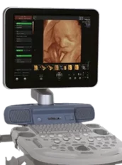
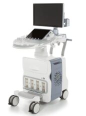
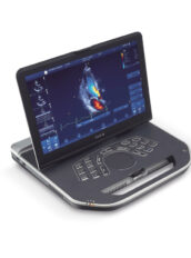
GE Voluson S10 FAQs:
Auto Optimization is a one-touch image optimization function that a user to optimize the image based on the actual B-mode image or Pulse wave Doppler data. The function works based on preset levels (Low, Medium, and High) and allows user to pick a preference for the contrast enhancement in the resulting image. Low does the least amount of contrast enhancement, high does the most. Auto Optimization is available in single or multi image, on live, frozen or CINE images (in B-Mode only), while in zoom, and in Spectral Doppler. Auto Optimization in PW Doppler Mode optimizes the spectral data. Auto adjusts the Velocity Scale (live imaging only), baseline shift, dynamic range, and invert (if preset). Upon deactivation, the spectrum is still optimized.
CrossBeam (Spatial Compounding Imaging) obtains real time sonographic information from several different angles of insonation and combines them to produce a single image. CrossBeam helps reducing speckle artifacts, enhancing mass margin delineation, and improving anatomical details.
SRI (Speckle Reduction Imaging) reduces speckle noise in images affects edges and fine details which limit the contrast resolution and make diagnostic more difficult.
Raw Data is a software tool that enables image processing, quick data re-acquisition, and image analysis with same resolution and same frame rates of original images. Raw Data helps shorten exam duration, improves clinical work flow by post-processing, and reduces a time to put probe on a patient.
Auto IMT automates measurement of intima-media thickness of vessel. Auto IMT helps keep tracking atherosclerosis diseases from the early stage as it is developed.
Scan Assistant is the customable scanning assistant tool that enables a user to save scanning protocols of each lab that help decrease key stokes up to 60~70% of ordinary operation. With fewer user’s action, an exam can be complete because a user does not need to move all around console and requires only one button push at most of exam.
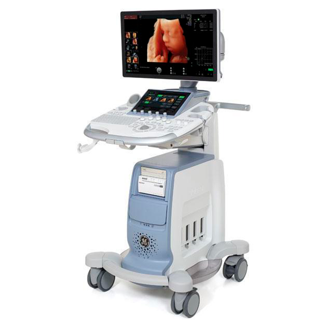
HDlive provides a movable virtual light source and calculates the propagation of light through skin and tissue. The user can freely position the light at any angle relative to the ultrasound volume to illuminate areas of interest. HDlive helps increase depth perception, reveal hidden details and can provide a deeper understanding of relational anatomy.
SonoBiometry – Performs a semi-automatic measurement of the head both head circumference and bi-parietal diameter), abdomen and femur. This tool can help enhance clinical workflow through helping reduce
Key strokes to perform biometry measurements.
SonoNT (Sonography-based Nuchal Translucency) and SonoIT (Sonography-based Intracranial Translucency) – Voluson technologies that help provide semi-automatic, standardized measurements of the nuchal and intracranial translucency as early as 11 weeks. Both tools can integrate easily into your workflow. SonoNT helps avoid the inter-and intra-observer variability that comes with manual measurements, and helps provide you with the reproducibility you demand.
SonoAVC*follicle (Sonography-based Automated Volume Count follicle) – Automatically calculates the number and volume of hypoechoic structures in a volume sweep, helping improve efficiency and workflow of follicular assessment. This feature helps to detect low echogenic objects (eg. follicles) in an organ (eg. ovary) and analyzes their shape and volume. From the calculated volume of the object an average diameter will be calculated. All objects detected that way will be listed according to size. The calculation results are displayed in the right monitor area. The objects are listed according to size. All different objects are color coded i.e. the color surrounding the number of the object also denotes the object on the image. If the mouse cursor hovers over a specific item on the list the respective object in the image is highlighted and vice versa. The color of the object is bound to its position on the list.
SonoVCADlabor – Helps you evaluate second-stage labor progression, and automatically documents the labor procedure with objective ultrasound data.
Advanced STIC (Spatio-Temporal Image Correlation) enables detailed three-dimensional images of the heart that can be viewed in motion. Clinicians can visualize an entire fetal heart cycle information that can help improve detection of heart defects with confidence
SonoVCAD*heart (Sonography-based Volume Computer Aided Display heart) – Helps standardize image orientation of the fetal heart by providing views automatically obtained from a single volume acquisition.
Advanced STIC (Spatio-Temporal Image Correlation) enables detailed three-dimensional images of the heart that can be viewed in motion. Clinicians can visualize an entire fetal heart cycle information that can help improve detection of heart defects with confidence
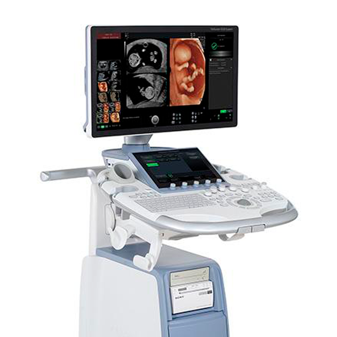
Tomographic Ultrasound Imaging (TUI) is a “Visualization” mode for 3D and 4D data sets. The data is presented as slices through the data set, which are parallel to each other. An overview image, which is orthogonal to the parallel slices, shows the parts of the volume, which are displayed in the parallel planes. This method of visualization is consistent with the way other medical systems such as CT or MRI, present the data. The distance between the parallel planes can be adjusted to fit the requirements of the given data set. In addition it is possible to set the number of planes.
SonoVCAD*heart (Sonography-based Volume Computer Aided Display heart) – Helps standardize image orientation of the fetal heart by providing views automatically obtained from a single volume acquisition.
B-Flow utilizes gray scale imaging to visualize a blood flow with different gray intensities according to the reflectors speed and hemodynamics. B-Flow is less dependency on the user or scanning angle, but Color Doppler heavily dependent on scanning angle, and also provides higher frame rate and spatial resolution than Color Flow. B-Flow may help visualize vessel-wall irregularities, kidney perfusion, liver and spleen vasculature, and bladder reflux or jets.
Strain Elastography is a non-invasive diagnostic technique displaying the relative elasticity of tissue stiffness compared to surrounding tissue by a real-time color map, superimposed on conventional gray scale image. Most malignant lesions have a harder or stiffer consistency than surrounding benign tissue. This change in stiffness can also be present in chronic or inflammatory diseases. To displace the underlying anatomical structures, Strain Elastography requires manual palpation by the user, or compression or decompression of the target produced by the patient respiration. It can be an efficient tool to improve biopsy targeting, image and evaluate breast and testicular lesions, and perform a quick monitoring and follow up of interventional procedures. Additionally, it can provide additional information to increase diagnostic confidence for musculoskeletal diseases such as tendinopathies, tendinosis, synovial hypertrophy or tears.
Scan Assistant is the customable scanning assistant tool that enables a user to save scanning protocols of each lab that help decrease key stokes up to 60~70% of ordinary operation. With fewer user’s action, an exam can be complete because a user does not need to move all around console and requires only one button push at most of exam.

Reviews
There are no reviews yet.